Home Page Strabismus Course Strabismus Books Strabismus Videos

Strabismus Lectures Case of the week E Consultation Contact me


Case of the week 102  ( March 2013 )
Right Superior Rectus Palsy


The parents of this 13 years old boy were referred by an orthopediatrist searching for an ocular cause to the abnormal head posture of their son. They claim that this posture was noticed since early childhood.


An isolated paralysis of the SR muscle is most commonly of congenital origin. It may also be secondary to trauma, for instance, after the historical bridle suture during cataract surgery , in which instance the paralysis is usually temporary.

In SR palsy the primary defect is limited elevation in abduction. Elevation is normal in adduction. However, when superior rectus palsy has been present for long periods, elevation from primary position and adduction may also become limited simulating double elevator palsy ( pic 1 & 3 ) . The ipsilateral inferior rectus ( pic 7 ) and the contralateral inferior oblique ( pic 1 ) muscles overact, and a small 
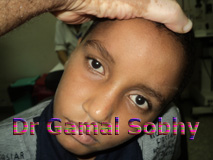
excyclotropia usually is present. The paralyzed eye is hypotropic in primary position ( pic 5 ) , and Bell’s phenomenon is absent ( In supranuclear DEP there is intact Bell’s phenomenon )

Abnormal Head Posture occurs frequently, but the position of the head is of little diagnostic significance in cases of SR palsy . Even though in most patients the head is tilted toward the sound side, the opposite may occur. In persons with this type of muscle paralysis of recent onset, the face is turned upward, the chin is elevated, and the head usually is inclined toward the sound side , the opposite may occurs .

Superior rectus muscle paralysis is frequently but not always associated with weakness of the homolateral levator palpebrae muscle ( pic 5 ) , particularly if the paralysis is congenital. A true ptosis caused by levator weakness must be differentiated from pseudoptosis secondary to the hypotropic position of the globe.

This patient with paralysis of the right superior rectus and  minimal ptosis of the right eye lid with the nonparetic eye fixating. When the paralyzed eye fixated, the ptosis disappeared and a marked hypertropia of the nonparalyzed eye occurred ( pic Lt head tilt ) .

The differential diagnosis of a superior rectus muscle paralysis includes mechanical causes that limit elevation of the eye, such as contracture, fibrosis, high myopia (heavy eye), myositis, endocrine orbitopathy, or a blow-out fracture of the orbital floor. Whenever elevation is restricted mechanically the forced duction test will be positive, and the restriction often involves the whole entire upper field of gaze.

For paralysis of the superior rectus muscle , a simple resection-recession operation of the vertical rectus muscles usually is effective. If the deviation is limited to upward gaze , 4-mm resection of the paretic muscle without recession of its antagonist may sufficient . The question arises whether surgery should be performed on the fixating or nonfixating eye. With rare exceptions, I prefer to operate on the paretic eye regardless of whether it is the dominant or nondominant eye. The amount of surgery that is necessary varies, of course, depending on whether the paretic eye (secondary deviation) or the nonparetic eye (primary deviation) habitually fixates.


الموقع المصري للحول وامراض الجهاز الحركي للعين

The Egyptian Site of Strabismus & Oculomotor Disorders