Case of the week 183 (
May 2015 ) Third Nerve Palsy
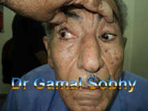
68 years old patient has had a sudden onset
of dropped right upper eyelid. The condition
started 2 months ago, the Rt eye
is deviated
outward and downward. Because of the ptosed led,
the patient has double vision on
elevating the dropped eyelid.
He is diabetic and
hypertensive. The pupil of the Rt eye is semidilated not reactive. Fundus examination shows normal
fundus. CT brain and orbit shows
no significant data
4 months later
After 12 months
See video of this patient
Third Nerve Palsy
Third nerve palsy, whether complete ( involving
both the intrinsic muscles of the eye together
with the extraocular muscles ) or
incomplete ( affecting only the extraocular
muscles ) has different
outcomes.
1. Complete recovery may occur soon in a week or
two. Rapid recovery suggests neurapraxia, without axonal
loss.
2.
In diabetics or hypertensive patients, the
pathology is vascular where nerve infarctions
occur and recovery does not begin for a month or more but is
usually complete within 3 months . This suggests a
lesion of axons, with preservation of nerve
connective tissue.
3.
With aneurysmal compression or
trauma, it can take up to 2 or 3 years to recover
completely, suggesting more severe anatomic
disruption of the nerve. If ptosis following trauma
is going to recover completely, it usually does so
within 6 months but occasionally takes as long as 2
years
4.
In some cases of third nerve palsy, the
paralysis persists completely unchanged. These
nerves have usually been transected by trauma or
chronic compression or have been infiltrated by
tumor.
5. Sometimes recovery is only partial. This
occurs especially after damage to the fascicular
portion of the nerve. Partial recovery may be
characterized by oculomotor nerve synkinesis,
so-called aberrant regeneration. Usually this
synkinesis becomes apparent within 9 weeks after
injury, but it has taken 3 to 6 months
This 68 years old diabetic and hypertensive patient has complete third
nerve palsy 2 months ago , most probably of
vascular origin , no history of trauma ,
normal fundus with bilateral normal optic nerve head. I usually ask for brain MRI
in acquired cranial nerve palsy to rule out
compressive lesions.
The patient was followed up at 4 months
interval, after 1 year he showed more or less
complete recovery of all ocular motility. He
still has right XT which may be also recovered
later spontaneously.
الموقع المص��ي ������ل����ول
وامراض الجهاز الحركي للعين
The Egyptian Site of
Strabismus & Oculomotor Disorders
.jpg)