The initial step in the
evaluation of orbital disease is a complete
ophthalmic examination. A careful medical and
ophthalmic history, including time course of the
disease, past trauma, ocular surgery, and
systemic illnesses, must be obtained. A complete
clinical examination includes assessment of
visual acuity and visual fields, anterior and
posterior segment evaluation, and external and
periorbital inspection. The use of modern
imaging techniques is almost always indicated -
the choice depends on the disease processes
suspected.
Osteomas are the most common
fibro-osseous lesions in the paranasal sinus.
They are benign tumours characterized by slow
growth and may extend to surrounding
structures.
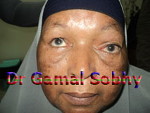
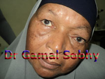
The patients usually
presented with severe inferior lateral
displacement of the eye ball, superior
limitation of ocular motility and sometimes proptosis were detected.
Anteroposterior x-ray of
the skull showed a large, dense, mushroom shaped
mass projecting into the orbit from the orbital
roof.
Axial and coronal computed tomography images
revealed a well-defined, multilobulated high
density tumor, orginated from the superior wall
of the frontal sinus and involving the majority
of the anterior superior orbital region.
Extension into the orbit with narrow neck was
noted.
The choice of surgical
management depends on the location, size and
experience of the surgeon. An open approach
allows tumor removal with direct visual control
and remains the best option in large tumors, but
the continued progression in endoscopic
approaches is responsible for new indications in
closed techniques. Immediate reconstruction
allows aesthetic and functional restoration of
neighbouring structures, which should one of the
goals in the treatment of this benign entity.
.jpg)