|
The
difficulty encountered by the clinician in
diagnosing a superior oblique palsy in view
of its being confused with a superior rectus
palsy of the fellow eye is a common finding in
ocular motility examination.
A contracture of the
antagonist
of the paralyzed left SO
muscle ( which is the left IO ) not only obscure
the nature of the primary defect in the
paralyzed eye but also affect motor balance of
the fellow eye when the patient fixates with the
paralyzed eye. The left IO muscle will require
less innervation to move the eye in its field of
action since the normal tonus of its paralyzed
opponentis decreased. According to Hering’s law
of equal innervation, the yoke muscle of the
antagonist ( the right SR muscle ) will receive
less innervation than required and will be
underacting ( Picture 1 ). This phenomenon is called
( inhibitional
palsy of the contralateral antagonist )
and it presents difficulties in diagnosis. Like
here in our patient who has a a left superior
oblique palsy who habitually in whom the right
superior rectus appears to be paretic (
the right eye is hypotropic and consequently the
right eyelid appears slightly ptosis, a picture
may be confused with right SR palsy. The
differentiation between the two conditions is
based upon ocular motility findings and on
Bielschowsky head tilt test.
The
diagnostic and clinical features in this patient
with a left superior oblique palsy are :
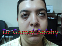
The head is
tilted to the right shoulder and the face is
turned to the right
( top picture ). In primary position
there is left hypertropia of 20 prism diopters (
Picture 5 ),
increasing to 30 prism diopters in adduction (
Picture 4 ), with the greatest
deviation (40 prism diopters) when the patient
is looking up and to the right
( Picture 1 ).
The hyperdeviation is also present in the left
field of gaze
where it
measured 10 prism diopters (spread of comitance,
Picture 6 ).
There is secondary overaction of the left
inferior oblique muscle
and limitation of
depression when looking down and to the right
( Picture 7 ).
The Bielschowsky head tilt test is diagnostic
for a left superior oblique paralysis with
increase of the left hypertropia on tilting the
head to the left shoulder and nearly no
hypertropia on
ilting the head to the right shoulder.
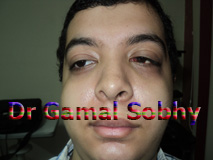
The plan of surgery of this
patient is to perform left IO myectomy ( with or
without right IR recession ), here is our
patient 10 hours post operative ( only IO myectomy was done )
|
.jpg)