Duane syndrome was described before the turn of
the 20th century. It is characterized by
limitation of abduction or adduction, narrowing
of the fissure with enophthalmos, up/down shoot
and face turn. All of these features vary
according to the class of Duane.
The etiology of Duane syndrome is misdirection
of the third nerve innervation to the medial
rectus. This misdirected medial rectus
innervation goes to the lateral rectus in the
orbit.
Co-contraction of the medial and lateral recti
is evident in electromyography.
Duane Type I is the most common (78%), followed by type III (15%) and II (7%).
Children with Duane syndrome
rarely complain. Parents usually bring their
children for examination because of strabismus
in the primary position
or because of the face turn. Parents
are usually unsure about the specific problem.
They often said that there is ‘something wrong.’
On the other hand, adults with Duane syndrome
often complain of asthenopia, intermittent
diplopia, and face turn.
Indications for surgery for Duane syndrome are :
ET/XT in the primary position, unacceptable head
posture, severe up- and downshoot, and
disfiguring enophthalmos.
The patient must be informed
that there is no surgery for Duane that will
restore normal ocular comitance in all gaze
positions.
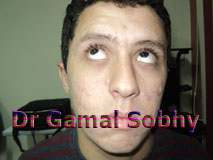
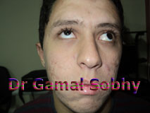
This patient has
Duane retraction syndrome Type III which is
characterized by the following features:
1. Congenital onset.
2. Limitation of
abduction
3. Limitation of
adduction
4. Globe retraction and
narrowing of the palpebral fissure on adduction
5. Upshoot in adduction
6. XT in the 1ry position ( Exotropic Duane )
This patient fulfils all indications of surgical
interference in Duane Syndrome.
For exotropia
associated with the
retraction syndrome, recession
of the lateral
rectus muscle of the involved eye is recommended.
For Narrowing of the
fissure, recession of the medial rectus combined
with recession of the lateral rectus is
recommended.
For the uphoot on
adduction, Y splitting of the lateral rectus is
recommended.
Many authors
reported good results
with a posterior
fixation suture
of the horizontal rectus muscles of the sound
eye to lessen the excursions of the sound eye on
looking nasally and temporally.
.jpg)